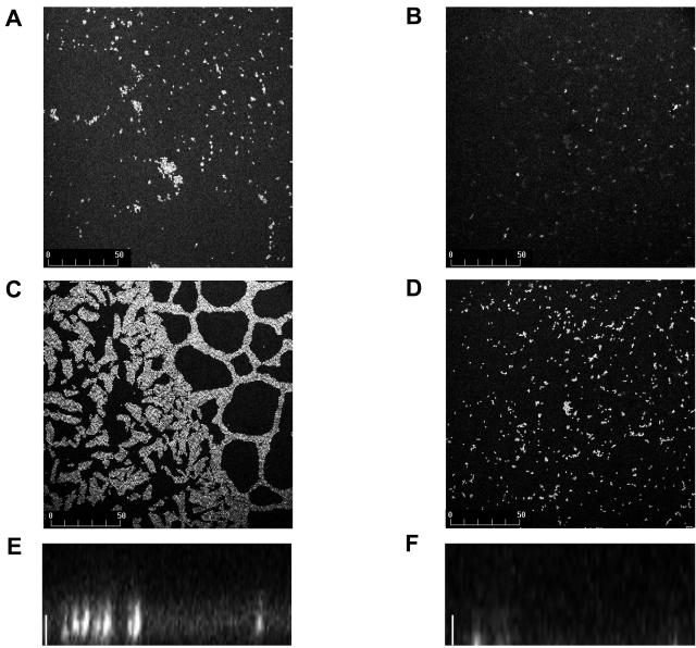
FIG. 3.
Confocal laser scanning microscopy of E. faecalis biofilms. The 24-h (A and B) and 48-h (C and D) biofilms were stained with acridine orange and visualized by confocal laser scanning microscopy as described in Materials and Methods. Standard projections of the biofilm through the x-y plane for strain V583 (A and C) and strain JML101 (fsrA) (B and D) are shown. Bars (A to D) indicate size in microns. Projections of the biofilms through the x-z plane at 48 h for strains V583 (E) and JML101 (fsrA) (F) are also shown. Bars (E and F), 10 μm.

