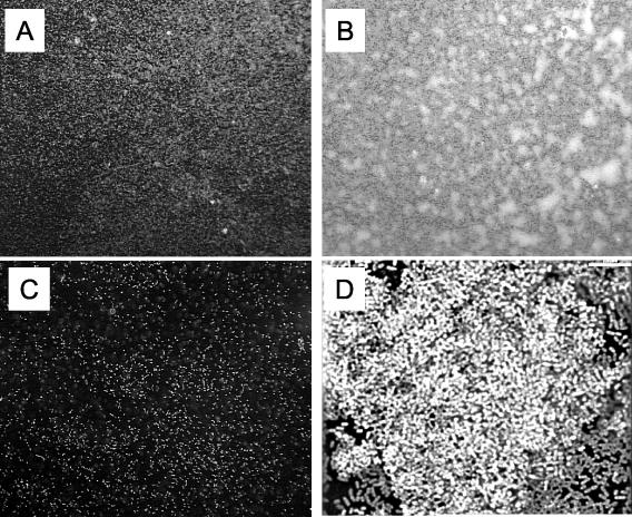
FIG. 2.
Formation of microcolonies by B. bronchiseptica on glass coverslips. Wild-type B. bronchiseptica organisms were grown on glass coverslips and then were stained with Syto Red 17 and observed under a fluorescent microscope (20× objective and 10× eyepiece). (A) Culture medium with no nicotinic acid (Bvg+ phase). (B) Culture medium supplemented with 0.8 mM nicotinic acid (Bvgi phase). (C) Culture medium supplemented with 4 mM nicotinic acid (Bvg− phase). Bacteria grown in Bvg+ phase (A) appear to form small aggregates, whereas microcolonies formed by bacteria grown in Bvgi phase are large and distinct (B). Bacteria in Bvg− phase (C) displayed little adherence to the coverslip with no aggregative properties. (D) Deconvolution micrograph of a microcolony depicted in panel B displaying the cellular architecture of the microcolony. Bar, 7 μm.

