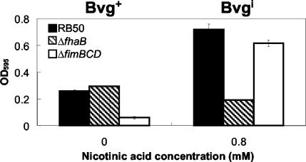
FIG. 4.
Quantitative assay of biofilm formation in wild-type B. bronchiseptica (RB50), ΔfhaB mutant, and ΔfimBCD mutant in the Bvg+ phase (0 mM nicotinic acid) and Bvgi phase (0.8 mM nicotinic acid). Bacteria were grown in 96-well polystyrene plates, and biofilm formation was quantified by absorbance of solubilized crystal violet stains, as described in Materials and Methods. The amount of biofilm formed by the ΔfhaB mutant in the Bvg+ phase was similar to that of the wild-type but was significantly decreased in the Bvgi phase. The ΔfimBCD mutant appears to form almost no biofilm in the Bvg+ phase, but the amount of biofilm formed by this mutant in the Bvgi phase was comparable to that of the wild-type bacteria. OD595, optical density at 595 nm.

