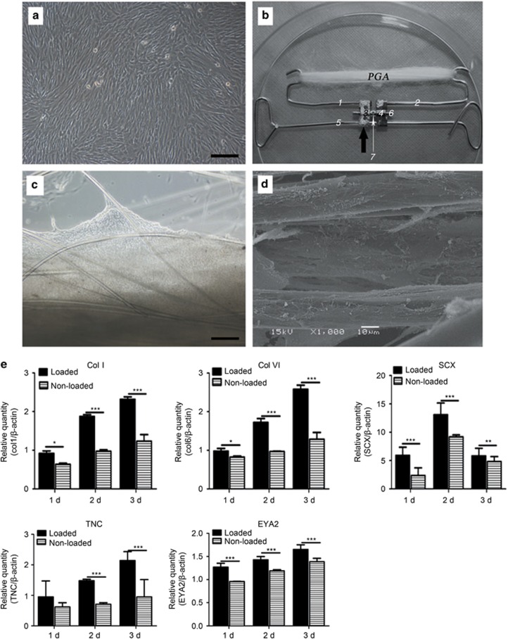
Figure 2.
In vitro culture of cell scaffold constructs. Design of tendon complex scaffold. (a) Passage 3 DPSCs cultured for confluence. (b) PGA long fibres were hooked on a custom-fabricated spring. Dark arrow points to a commercial dental arch expansion appliance (including flanks 3 and 4 and central screw axis 6). Wires 1 and 2 are fused onto flanks 3 and 4, respectively. Flanks 3 and 4 can be finely expanded along screw axis 6 by turning a hole in it, and these three parts as a whole can slide along wire 5; the latter constrains the entire appliance in the cell culture dish using two loops. (c) Phase-contrast microscopy of a cell-scaffold construct cultured for 1 week. (d) SEM view shows attachment and spreading of DPSCs on PGA fibres and their matrix production. Original magnifications: (a, c) × 100; bar=200 μm, (d) × 1000, bar=10 μm. (e) Quantitative real-time PCR confirms the gene expression of tendon-specific and tendon-related matrix genes in engineered tendon scaffolds with or without loading after 1–3 days (n=3, mean±SD). Gene expression levels are normalized to the reference gene β-actin (y axis); the mRNA harvested from DPSCs was cultured in a dish as a control group. *P<0.05 between two groups; **P<0.01 between two groups; ***P<0.001 between two groups (P-values are provided in Supplementary Data S3). DPSC, dental pulp stem cell; PCR, polymerase chain reaction; PGA, polyglycolic acid; SD, standard deviation; SEM, scanning electron microscope.

