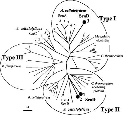
FIG. 2.
Phylogenetic analysis of the A. cellulolyticus ScaD cohesins. The first two ScaD cohesins map together with the ScaB cohesins on a separate branch of the type II cohesins, radiating from approximately the same bifurcation point. In contrast, the third ScaD cohesin is clearly a member of the type I cohesins, closely aligned to the seven type I cohesins of ScaA. The scale bar indicates the percentage (0.1) of amino acid substitutions. Sources for sequences used in this figure are provided in references 15, 16, 46, and 56.

