Figure 1.

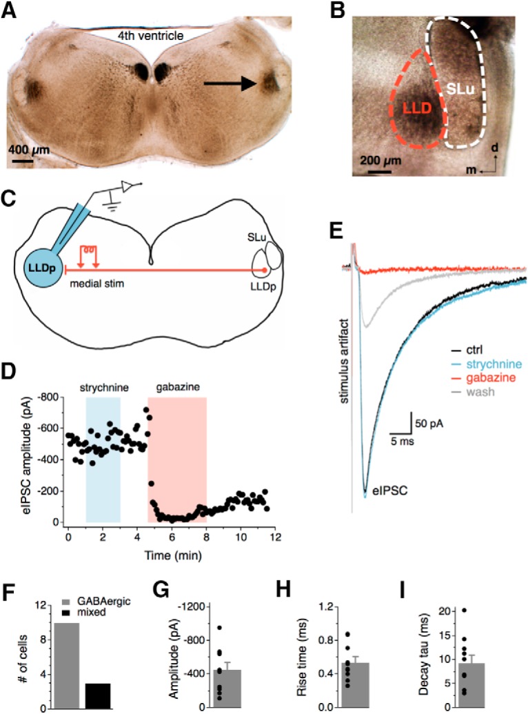
Synaptic inhibition in the LLDp is largely GABAergic. A, The LLDp (arrow) is located laterally within a 300-μm-thick coronal brainstem slice with ∼4 mm between the two LLDps. B, The LLDp is readily identifiable as a heavily myelinated nucleus medial and slightly ventral to the semilunar nucleus (SLu) in fresh tissue slices. C, Schematic of the experimental setup highlighting ipsilateral recording site (blue, left), and medial electrical stimulation of fibers projecting from the contralateral LLDp. D, Bath application of gabazine (10 μm), a GABAA receptor antagonist, abolished the eIPSC, whereas the eIPSC amplitude was not affected by strychnine (1 μm), a glycine receptor antagonist. E, Averaged traces of the eIPSC during control (black), strychnine (1 μm, blue), gabazine (10 μm, red), and wash (gray). F, In the majority of cells, eIPSCs were abolished by gabazine alone (n = 10), although occasionally an additional weak strychnine component was observed (n = 3). G–I, Population data of eIPSC amplitude, 20–80% rise time, and decay time constant (tau) for GABAergic eIPSCs (n = 10). For this and subsequent figures, mean ± SEM values are shown. d, dorsal; m, medial; ctrl, Control; stim, stimulation.
