Figure 2.
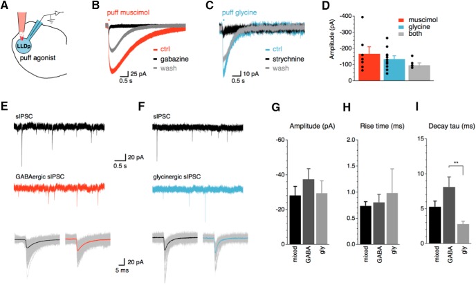
LLDp neurons have functional GABAA and glycine receptors. A, Schematic of the experimental setup highlighting direct puff application of receptor agonists to the recorded cell. B, C, In all neurons recorded, puff application of muscimol (GABAA receptor agonist, 10 μm, red; B) and glycine (glycine receptor agonist, 500 μm, blue; C) evoked IPSCs, which were blocked by their respective antagonists, gabazine (10 μm) and strychnine (1 μm). D, Amplitude of IPSCs did not significantly differ among puff application of muscimol (n = 7), glycine (n = 11), or a mixture of muscimol and glycine (n = 5), suggesting that GABA and glycine may interfere with each other. E, F, sIPSCs (top, black) were pharmacologically isolated into GABAergic sIPSCs (E, red) or glycinergic sIPSCs (F, blue) with bath application of strychnine (1 μm) or gabazine (10 μm), respectively. Averaged sIPSCs (bottom, thick lines) show distinct decay kinetics between GABAergic and glycinergic sIPSCs. G–I, Population data of sIPSC amplitude, 20–80% rise time, and decay tau. Population data for decay tau (I) shows significant difference between GABAergic (n = 3) and glycinergic sIPSCs (n = 3; p = 0.008, ANOVA with post hoc Fisher’s exact test). For this and subsequent figures: *p < 0.05, **p < 0.01, and ***p < 0.001. ctrl, Control; gly, glycine.

