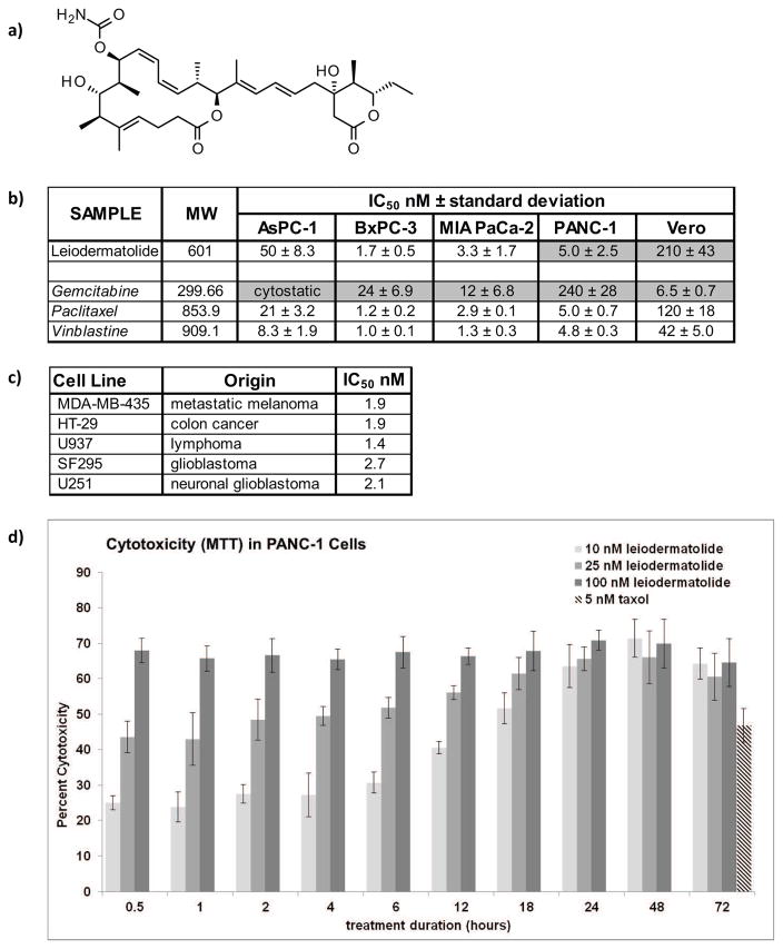
Figure 1. Structure of Leiodermatolide and Induction of Cytotoxicity in Cancer Cells.
a) Structure of Leiodermatolide. b) Four different pancreatic cancer cell lines were treated for 72 hours in the presence of vehicle controls, media alone, and a range of concentrations of leiodermatolide, paclitaxel and vinblastine. Changes in color due to metabolization of MTT by live cells were measured, normalized against vehicle control and the IC50 determined using a non-linear regression curve fit. The resulting average of 3 experiments ± standard deviation is shown. The IC50 of leiodermatolide for the PANC-1 and Vero cell lines13, and the IC50 for Gemcitabine31 in these pancreatic cancer cell lines (shaded) have been previously reported and are provided here for ease of comparison. Leiodermatolide caused cytotoxicity to pancreatic cancer cells in low nanomolar concentrations. A much higher concentration was needed to cause similar cytotoxicity in the non-carcinogenic Vero cell line. Leiodermatolide exhibits similar potency to paclitaxel and vincristine with better selectivity, and more potency and selectivity than gemcitabine. c) Leiodermatolide induces potent cytotoxicity in other cancer cell lines of different origin. d) Washout experiments were conducted using 10, 25, 100 nM leiodermatolide in PANC-1 cells. Treatments were removed by aspiration at the times listed and replaced with pre-warmed media. Cytotoxicity was measured at 72 hours. Paclitaxel was used as a control at its IC50 as a positive control. Only treatments with 10 nM leiodermatolide were reversible if the compound was removed within 12 hours. The average of 3 experiments ± standard deviation is shown.

