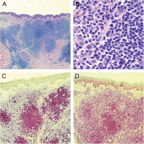
Fig 1.

Histological findings of the skin biopsy from initial presentation. a Low magnification (25×) demonstrating a nodular infiltrate in the superficial and deeper dermis (H&E). b At higher power (400×) the majority of the cells are small and have irregular nuclei and inconspicuous nucleoli. There are scattered large transformed immunoblasts (H&E). c CD20 stain highlights the nodules of B lymphocytes (CD20 immunohistochemical stain, 50×) d The B lymphocytes in the nodules are positive for BCL2 (BCL2 immunohistochemical stain, 50×)
