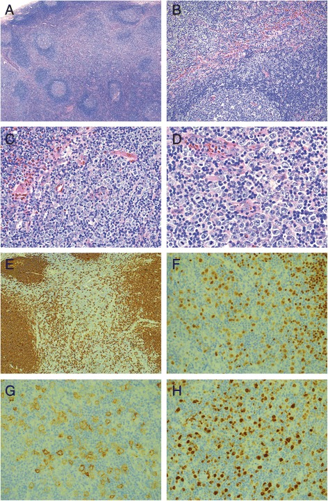
Fig. 2.

Histological findings of the inguinal lymph node excision 2 weeks after initial presentation. a Low-power view (25×) shows a lymph node with a preserved architecture with intact capsule, patent sinuses and polarized germinal centers with well-demarcated mantle zones. The interfollicular areas are expanded (H&E). b At higher magnification (100×) the interfollicular infiltrate is composed of numerous large transformed immunoblasts in the background of small lymphocytes, abundant eosinophils, scattered plasma cells and histiocytes (H&E). c At 200× magnification (H&E) and (D) at 400× magnification the large atypical cells have large oval to slightly irregular nuclei with vesicular chromatin, prominent nucleoli and scant to moderate amounts of clear to amphophilic cytoplasm (H&E). e The large atypical cells are immunoreactive for CD20 (CD20 immunostain, 50×) as well as for f PAX5 (PAX5 immunostain, 200×), g CD30 (CD30 immunostain, 200×) and h MUM1 (MUM1 immunostain, 200×)
