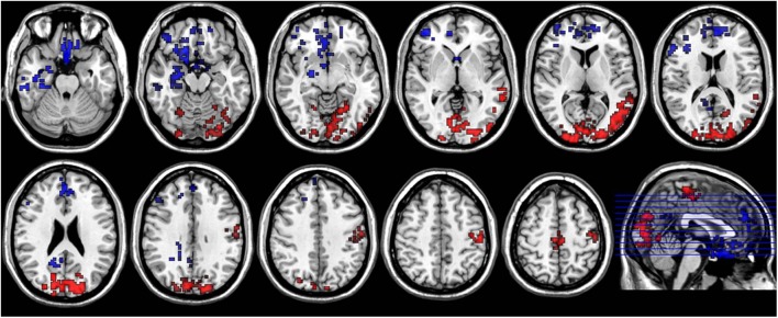
FIGURE 5.
Axial sections show areas with significant diurnal variations of both ReHo and ALFF. The left hand side of the image is the right side of the brain (radiology convention). The hot color indicates increased ReHo and ALFF in AM compared to PM, and cool color indicates decreased ReHo and ALFF.

