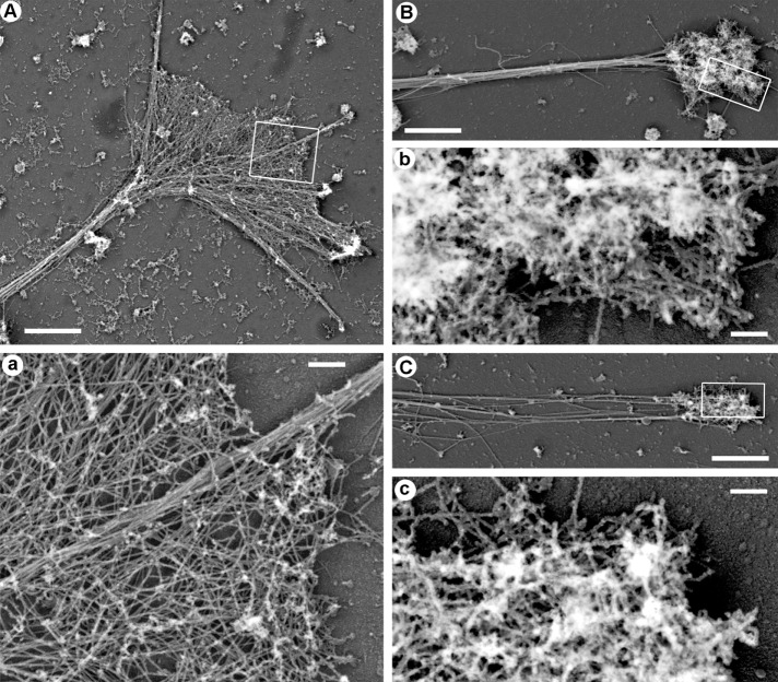
FIGURE 2:
PREM of cytoskeleton structure in distal neurite regions. (A) Growth cone and a distal portion of the neurite from a neuron cultured in a normal medium. (a) Boxed region in A enlarged to show actin filament bundle in the filopodium and branched actin network in lamellipodia. (B, C) Actin-rich bulbs and the distal neurite regions from neuron treated with 2.5 μM LatB (B) or 2.5 μM CytoD (C). (b, c) Boxed regions in B and C, respectively, enlarged to show dense branched actin network in the actin bulb. Scale bars, 2 μm (A–C), 200 nm (a–c).

