
FIGURE 3:
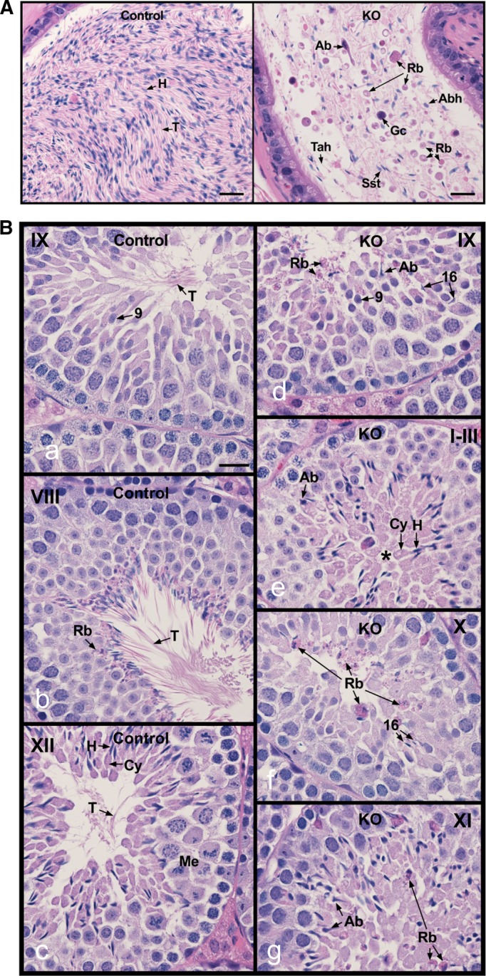
Histology of the cauda epididymis and testis from control and conditional Ift20 mutant mice. (A) Epididymis from control mice showing highly concentrated epididymal sperm with aligned sperm tails (T) and normal sperm heads (H; left). (B) Epididymis from IFT20 knockout mice (right), showing a very low concentration of sperm, with high incidence of sperm abnormalities (Ab), sloughed round germ cells (Gc), residual bodies (Rb), sperm with short tails (Sst), and sperm tails without heads (Tah). Bars, 20 mm. Testes from control (a–c) and IFT20 knockout mice (IFT20 KO; d–g). (a) Stage IX, showing normal step 9 spermatids with long tails (T) extending into the lumen. (b) Stage VIII, showing mature sperm with long tails (T) being released into the lumen. Residual bodies (Rb) of leftover cytoplasm are being formed and phagocytized by Sertoli cells. (c) Stage XII, showing spermatocytes in meiotic division (Me), step 12 elongating spermatids with head (H) of condensed chromatin, and bulges of cytoplasm (Cy). The spermatid tails (T) are long and extend into the lumen. (d) Stage IX, showing abnormal step 16 elongated spermatids (Ab) that were not released into the lumen and residual bodies (Rb) that are being sloughed into the lumen. (e) Stages I–III, showing abnormal elongated spermatids (Ab). The elongated spermatids show some normal heads (H) and cytoplasm (Cy), but their tails do not appear in the lumen (*). (f) Stage X, showing failure of spermiation, with step 16 spermatid heads remaining in the seminiferous epithelium. Residual bodies (Rb) remain at the lumen rather than being phagocytized. (g) Stage XI, showing abnormal elongating spermatids (Ab) and sloughed residual bodies (Rb) remaining from prior stages. Bars, 20 mm.