FIGURE 1:

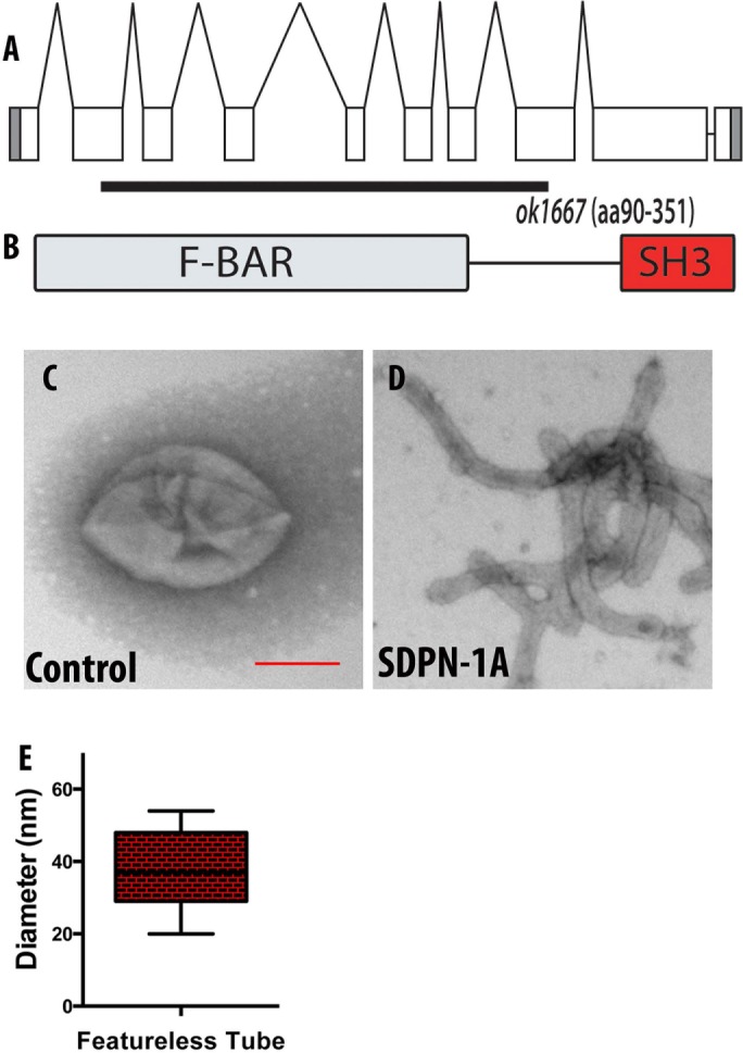
(A) Genomic structure of the sdpn-1 gene and the location of the ok1667 mutant deletion. ok1667 is a 2547–base pair deletion from the second exon to the eighth exon. (B) The ok1667 allele deletes sequences encoding amino acids 90–351, including the majority of the F-BAR domain, and places downstream sequences out of frame. (C–E) In vitro membrane tubulation of PtdSer liposomes by full-length SDPN-1a. Electron micrographs of acidic PtdSer liposomes (0.05 mg/ml, average diameter 400 nm) incubated with 2.5 μM (C) GST or (D) full-length SDPN-1. (E) Statistical analysis. Diameters of membrane tubules shown in D were quantified on at least three independently prepared electron microscopy grids. Scale bar, 200 nm.
