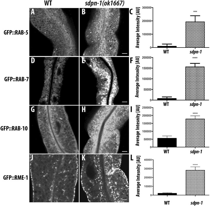
FIGURE 3:
sdpn-1 mutants affect early endosomes, basolateral recycling endosomes, and late endosomes. Laser scanning confocal micrographs of the worm intestine expressing GFP-tagged fusion proteins that are resident markers for distinct endosomal compartments. sdpn-1 mutants show abnormal intracellular accumulation of early endosomes labeled with GFP::RAB-5, late endosomes marked with GFP::RAB-7, GFP::RAB-10, which functions at the early endosome/basolateral recycling endosome interface, and basolateral recycling endosomes marked with GFP::RME-1. Control micrographs in the wild type background are shown for (A) GFP::RAB-5, (D) GFP::RAB-7, (G) GFP::RAB-10, and (J) GFP::RME-1. Confocal images in the sdpn-1(ok1667) mutant background are shown for (B) GFP::RAB-5, (E) GFP::RAB-7, (H) GFP::RAB-10, and (K) GFP::RME-1. Quantification of total fluorescence intensity for (C) GFP::RAB-5, (F) GFP::RAB-7, (I) GFP::RAB-10, and (L) GFP::RME-1. Error bars represent SEM: ***p < 0.001, ****p < 0.0001 (Student’s t test). Scale bar, 10 μm (B, E, H, K).

