FIGURE 6:
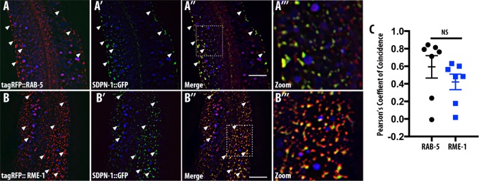
SDPN-1 resides on early and basolateral recycling endosomes. All micrographs are from deconvolved 3D confocal image stacks acquired in intact living animals expressing intestinal-specific GFP- and RFP-tagged proteins. (A–A′′′) SDPN-1::GFP colocalizes with RAB-5–labeled early endosomes. White arrowheads indicate endosomes labeled by both SDPN-1::GFP and tagRFP-RAB-5. (A′′′) Magnified image designated by the rectangular outline in A″. (B–B′′′) SDPN-1::GFP is also enriched on tagRFP-RME-1–labeled basolateral recycling endosomes. White arrowheads indicate positive colocalization between SDPN-1::GFP and tagRFP::RME-1. (B′′′) Magnified image designated by the rectangular outline in B″. (C) Pearson’s r for colocalization of SDPN-1::GFP with tagRFP-RAB-5 and tagRFP-RME-1. Six animals. Error bars represent SEM.

