FIGURE 1:
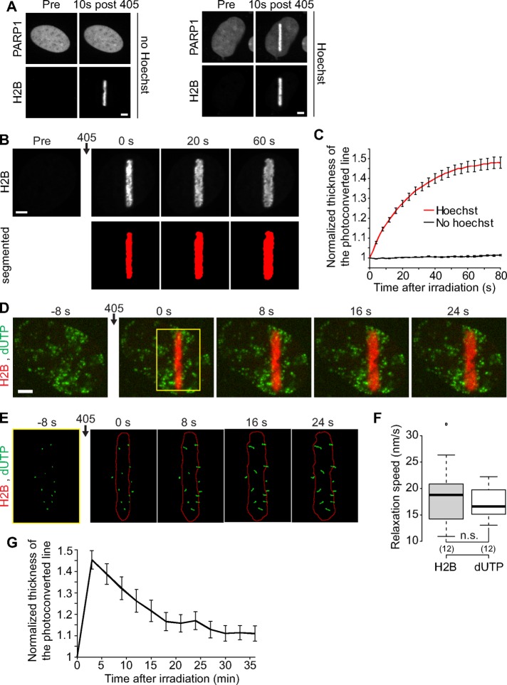
DNA damage induced by laser microirradiation induces transient chromatin relaxation. (A) Recruitment of PARP1 at the microirradiated area in cells coexpressing PARP1-mCherry and H2B-PAGFP. Scale bar: 4 μm. In cells not presensitized with Hoechst, the 405 nm irradiation induces local photoactivation of the H2B-PAGFP but no recruitment of PARP1-mCherry. In contrast, in the case of Hoechst presensitization, the 405 nm irradiation induces both photoactivation of the H2B-PAGFP and a marked recruitment of PARP1-mCherry, indicating the presence of DNA lesions. Similarly, we observed the recruitment of 53BP1 only in cells presensitized with Hoechst (unpublished data). (B) Confocal image sequence of a human U2OS nucleus expressing H2B-PAGFP. Scale bar: 4 μm. The automatic segmentation of the histone H2B channel is shown in red below the raw images. The average thickness of the segmented line can be plotted as a function of time after irradiation, as shown in C for cells presensitized (n = 17) or not (n = 23) with Hoechst (mean ± SEM). Based on this analysis, the ratio between the thicknesses of the photoconverted line at time = 60 s and time = 0 s can be calculated to estimate the relative relaxation of the irradiated region. (D) Confocal image sequence of a U2OS cell expressing H2B-PATagRFP (red) and labeled with fluorescent nucleotides dUTP-ATTO633 (green). Scale bar: 4 μm. (E) Enlarged view of the region overlaid in yellow on the previous panel. The segmentation of the photoconverted chromatin area (red outline) and trajectories of individual foci labeled with fluorescent nucleotides (green) are shown. For this experiment, the power of the 405 nm laser used for simultaneous photoactivation and microirradiation was set to 250 μW at the sample level, instead of 125 μW, to induce an enhanced chromatin relaxation, allowing an easier identification of the phase of directed motion for the dUTP-labeled foci. (F) Comparison between the speed at which the width of the H2B-labeled region is growing and the speed of the dUTP-labeled foci perpendicular to the irradiation line. We show the average speed for the 30 s subsequent to laser microirradiation. p Values were calculated by paired t test. (G) Dynamics of the chromatin compaction state at DNA damage sites over long timescales measured in wild-type U2OS cell expressing H2B-PATagRFP (mean ± SEM, n = 16).

