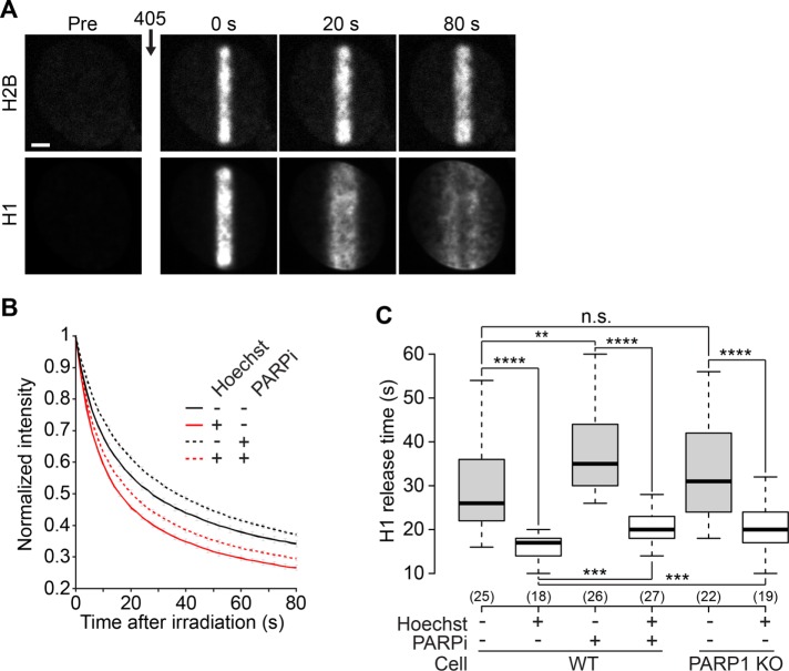
FIGURE 3:
The linker histone H1 is mobilized at DNA lesions. (A) Confocal image sequence of a U2OS nucleus coexpressing H2B-PATagRFP and H1.1-PAGFP. For the H1 channel, the image contrast was enhanced to allow the visualization of H1 redistribution over the entire nucleus following laser microirradiation. This led to an apparent saturation of the image at time = 0 s. Scale bar: 4 μm. (B) Kinetics of the release of the H1 proteins localized at the DNA lesions at the time of laser microirradiation in wild-type cells coexpressing H2B-PATagRFP and H1.1-PAGFP, presensitized or not with Hoechst and treated or not with the PARP1 inhibitor AG14361 (30 μM, 1 h; PARPi) (mean ± SEM, for each condition, 17 < n < 28). (C) Characteristic release time for H1, measured at half fluorescence decay, in wild-type and PARP1 KO cells.

