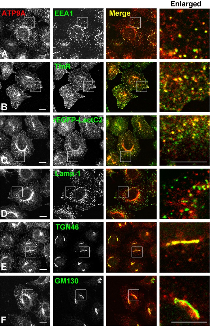
FIGURE 1:
Subcellular localization of ATP9A. HeLa cells stably expressing C-terminally HA-tagged ATP9A were doubly stained for HA and organelle markers EEA1 (early endosome), TfnR (early/recycling endosome), Lamp-1 (late endosome), TGN46 (TGN), and GM130 (cis-Golgi), followed by Cy3-conjugated anti-rat and Alexa Fluor 488–conjugated anti-mouse or anti-rabbit secondary antibodies. The recombinant EGFP-Lactadherin C2 domain (rEGFP-LactC2) was treated with anti-HA antibody after permeabilization of cells with digitonin. Images were obtained by confocal microscopy. Insets are enlarged in the right panels. Scale bars: 10 μm.

