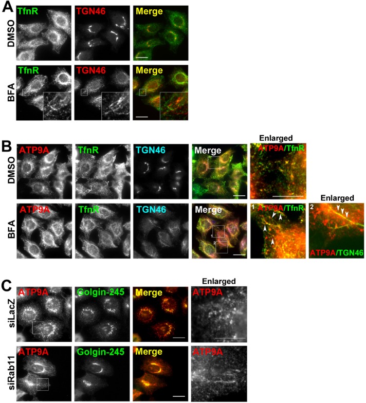
FIGURE 2:
Morphological changes in ATP9A-positive compartments following treatment with BFA or knockdown of Rab11. (A) HeLa cells or (B) HeLa cells stably expressing ATP9A-HA were treated with vehicle (dimethyl sulfoxide) or 5 μg/ml BFA for 15 min. (A) Cells were fixed and doubly stained for TfnR and TGN46, followed by Alexa Fluor 488–conjugated anti-mouse and Alexa Fluor 555–conjugated anti-rabbit secondary antibodies. (B) Cells were triply stained for HA, TfnR, and TGN46, followed by Cy3-conjugated anti-rat, Alexa Fluor 488–conjugated anti-mouse, and DyLight649-conjugated anti-rabbit secondary antibodies. Insets were enlarged. Scale bar: 10 μm. Arrowheads indicate colocalized tubular structures. (C) HeLa cells stably expressing ATP9A-HA were treated with siRNAs against LacZ (control) or Rab11a and Rab11b and doubly stained for HA and Golgin-245, followed by Cy3-conjugated anti-rat and Alexa Fluor 488–conjugated anti-mouse secondary antibodies. Scale bars: 20 μm. Insets were enlarged. Scale bar: 10 μm.

