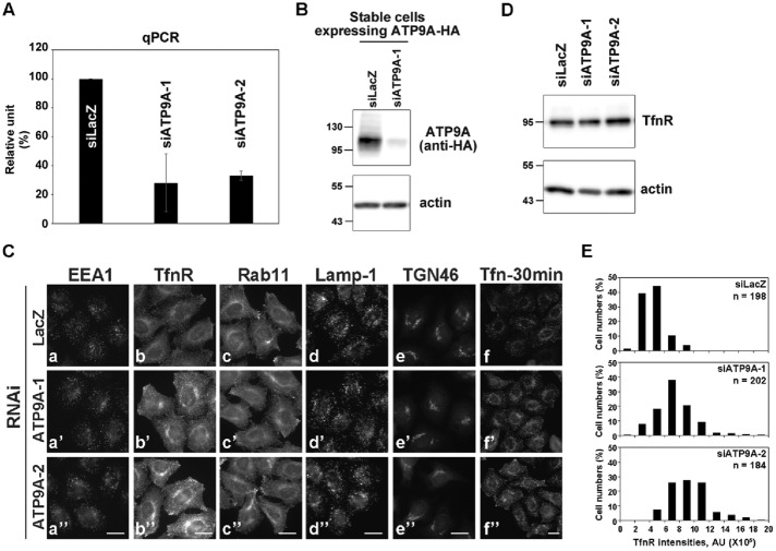
FIGURE 5:
Depletion of ATP9A does not affect the distribution of organelles. (A) HeLa cells were transfected with siRNAs against LacZ (control), ATP9A-1, or ATP9A-2, and total RNAs were isolated from the cells, and RT-qPCR was performed. The results are from three independent experiments (averages ± SD). (B) HeLa cells stably expressing ATP9A-HA were transfected with siRNAs against LacZ or ATP9A-1, and each cell lysate was analyzed by immunoblotting with anti-HA and anti-actin antibodies (as an internal control). (C) siRNA-treated HeLa cells described in A were stained for EEA1 (a), TfnR (b), Rab11 (c), Lamp-1 (d), or TGN46 (e). Cells were treated with Alexa Fluor 555–Tfn for 30 min at 37°C and then fixed (f). Scale bars: 20 μm. (D) siRNA-treated HeLa cells described in A were lysed, and the lysates were analyzed by immunoblotting with anti-TfnR and anti-actin antibodies. (E) Fluorescence intensities of TfnR in (C, f–f″) were quantitated using the MetaMorph software; the frequency distribution of intensities is shown.

