FIGURE 6:
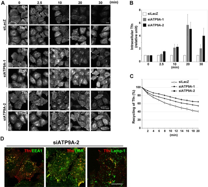
Depletion of ATP9A delays the recycling of Tfn. (A) HeLa cells were transfected with siRNAs against LacZ, ATP9A-1, or ATP9A-2; serum starved for 3 h; and then incubated at 4°C for 60 min with Alexa Fluor 555–conjugated Tfn. Cells were washed and incubated at 37°C for the indicated times and then fixed and stained for TfnR. Scale bars: 20 μm. (B) Pixel intensities of Alexa Fluor 555–conjugated Tfn were estimated at the indicated times using MetaMorph software. Data are shown as the ratio of the mean of cellular Tfn intensities between cells depleted of ATP9A and control cells at each time point. The graph is representative of three independent experiments, and 160–200 cells of each sample were analyzed. Graphs show means ± SE. (C) HeLa cells were transfected with siRNAs as described above, incubated at 37°C for 60 min with Alexa Fluor 555–conjugated Tfn, washed, and subjected to time-lapse recording. Images in multiple areas were captured every 2 min for a total of 20 min, and pixel intensities of Alexa Fluor 555–Tfn were quantitated in every image. Graphs show means ± SE from 25 cells for siLacZ, 86 cells for siATP9A-1, and 59 cells for siATP9A-2. Raw data are shown in Supplemental Figure S2. (D) HeLa cells treated with siRNA against ATP9A-2 were serum starved for 3 h, incubated at 4°C for 60 min with Alexa Fluor 555–conjugated Tfn, washed, incubated at 37°C for 20 min, and stained with anti-EEA1, anti-TfnR, and anti-Lamp-1 antibodies, followed by anti-mouse Alexa Fluor 488–conjugated secondary antibody. Images were obtained by confocal microscopy. Scale bars: 20 μm.

