
FIGURE 7:
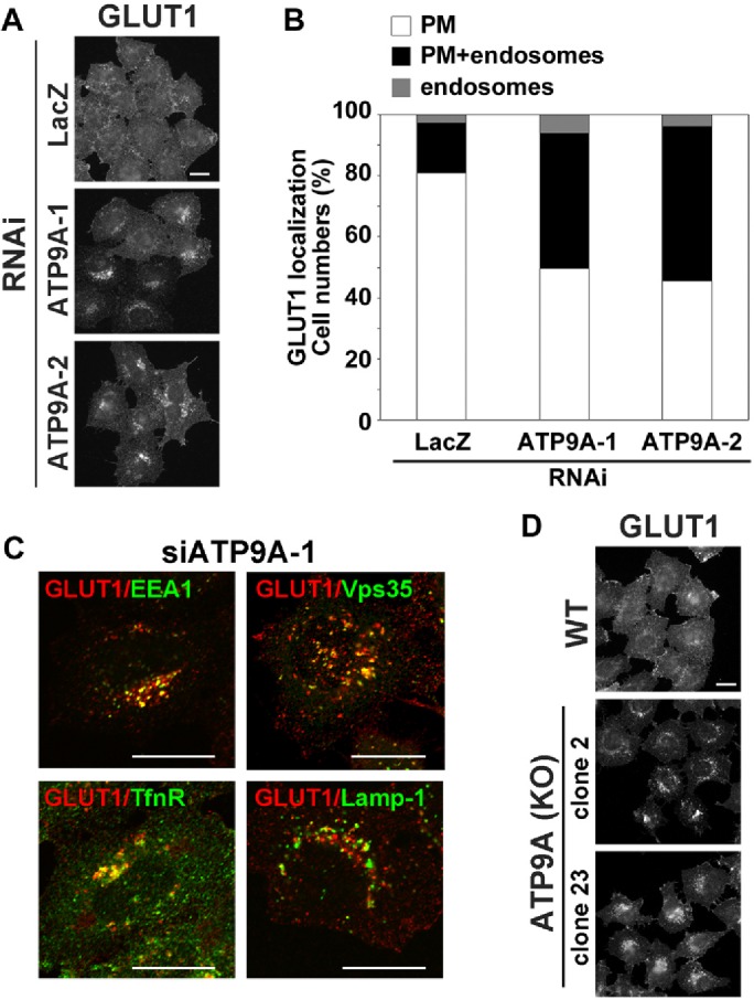
Depletion of ATP9A inhibits recycling of GLUT1. (A) HeLa cells were transfected with siRNAs against LacZ, ATP9A-1, or ATP9A-2, and then stained with anti-GLUT1 antibody. After being fixed, cells were incubated with Alexa Fluor–conjugated secondary antibody. Scale bar: 20 μm. (B) Cells in which GLUT1 localized to the plasma membrane (white), to the plasma membrane and endosomes (black), or to the endosomes (gray) in A were counted; counts were normalized against the total number of counted cells. A total of 214 cells for siLacZ, 199 cells for siATP9A-1, and 180 cells for siATP9A-2 were counted. (C) HeLa cells treated with siRNA against ATP9A-2 were fixed, permeabilized, and incubated with anti-GLUT1 and anti-EEA1, anti-Vps35, anti-TfnR, or anti-Lamp-1 antibodies, followed by Alexa Fluor 555–conjugated anti-rabbit and Alexa Fluor 488–conjugated anti-mouse secondary antibodies. Images were obtained by confocal microscopy. Scale bars: 20 μm. (D) Parental HeLa cells and two clones of ATP9A knockout cells were stained with anti-GLUT1 antibody. After fixation, cells were incubated with Alexa Fluor–conjugated secondary antibody. Scale bar: 20 μm.