FIGURE 1:
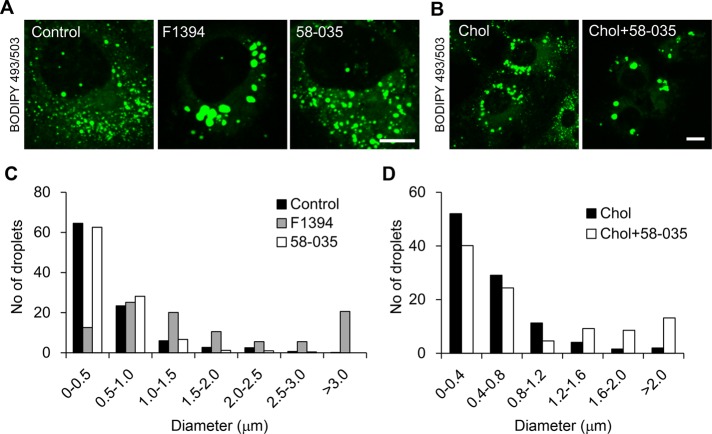
Treatment of Huh7 cells with ACAT inhibitors enlarges LDs. (A) Huh7 cells were cultured for 24 h in the absence (Control) or presence of 10 μg/ml F1394 or 10 μg/ml 58-035. Cells were then labeled with 2 μg/ml BODIPY 493/503 for 30 min. Bar, 10 μm. (B) Cells were treated with MβCD/cholesterol in LPDS (Chol) or MβCD/cholesterol plus 58-035 in LPDS (Chol + 58-035) as described in Materials and Methods. Cells were labeled with BODIPY 493/503. Bar, 10 μm. (C) The size distributions of LDs were quantified as described in Materials and Methods after 24-h treatment of Huh7 cells with 10 μg/ml F1394 or 10 μg/ml 58-035. (D) The size distributions of LDs were quantified after 24-h treatment of Huh7 cells with MβCD/cholesterol in LPDS (Chol) or MβCD/cholesterol plus 58-035 in LPDS (Chol + 58-035).

