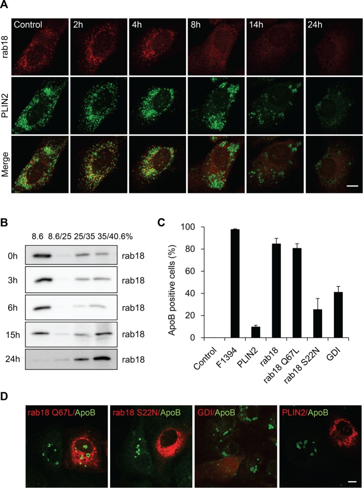
FIGURE 8:
Association of ApoB with LDs is dependent on the activity of Rab18. (A) F1394, 10 μg/ml, was added to Huh7 cells. At appropriate intervals, cells were fixed, permeabilized with digitonin, and doubly labeled with anti-PLIN2 and anti-Rab18 antibodies as described in Materials and Methods. Bar, 10 μm. (B) Cells were treated with F1394 as described and then homogenized, and PNS was separated by stepwise sucrose density gradient as described in Materials and Methods. The distribution of Rab18 was measured by Western blotting. (C, D) Huh7 cells overexpressing mCherry-PLIN2, mCherry-Rab18, mCherry-Rab18 S22N, mCherry-Rab18 Q67L, and mCherry-GDI were treated with F1394 as described in Materials and Methods, and the number of ApoB-positive cells was counted after immunofluorescence. Bar, 10 μm.

