FIGURE 7:
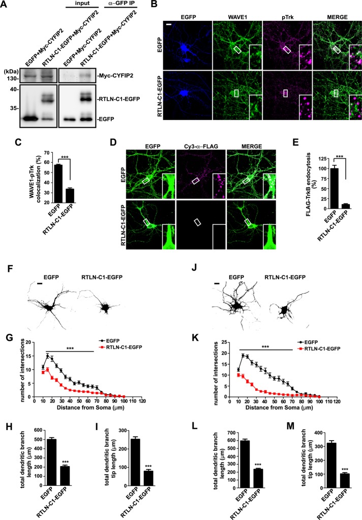
Disruption of the interaction between retrolinkin and CYFIP1/2 impairs BDNF-induced TrkB endocytosis and dendritic outgrowth. (A) Lysates from HEK 293T cells coexpressing Myc-tagged CYFIP2 and RTLN-C1-EGFP (shown in Figure 1A) were subjected to coIP assay with antibodies against GFP. Bound proteins were analyzed by SDS–PAGE and immunoblotting with antibodies against Myc and GFP. (B) Hippocampal neurons transfected with construct expressing EGFP or RTLN-C1-EGFP were starved for 2 h, stimulated with BDNF for 2 min, and immunostained with antibodies against WAVE1 and pTrk. (C) Quantification of the colocalization between WAVE1 and pTrk in B. Thirty neurons from three independent experiments were analyzed. Data represent mean ± SEM (N = 3). (D) Hippocampal neurons cotransfected with expression constructs for FLAG-TrkB and EGFP or RTLN-C1-EGFP were treated with BDNF (25 ng/ml) for 30 min. Internalized FLAG-TrkB was labeled with Cy3–α-FLAG. (E) Quantification of TrkB endocytosis in neurons in D by measuring mean fluorescence intensity of FLAG-TrkB puncta per neuron (35 neurons from three independent experiments). (F) Hippocampal neurons were transfected at DIV1 with EGFP or RTLN-C1-EGFP expression construct and fixed at DIV5, followed by immunostaining with anti-GFP. Scale bar, 10 μm. (G–I) Sholl analysis (G), quantification of total dendritic branch length (H), and total dendritic branch tip length (I) from neurons in F. Data from 30–35 neurons. All values are shown as mean ± SEM (N = 3). (J) Hippocampal neurons were transfected as described in F and cultured for 4 d in the presence of BDNF (25 ng/ml) after transfection. (K–M) Sholl analysis (K), quantification of total dendritic branch length (L), and total dendritic branch tip length (M) from neurons in J. Data from 30–35 neurons. All values are shown as mean ± SEM (N = 3). ***p < 0.001. Scale bar, 10 μm.

