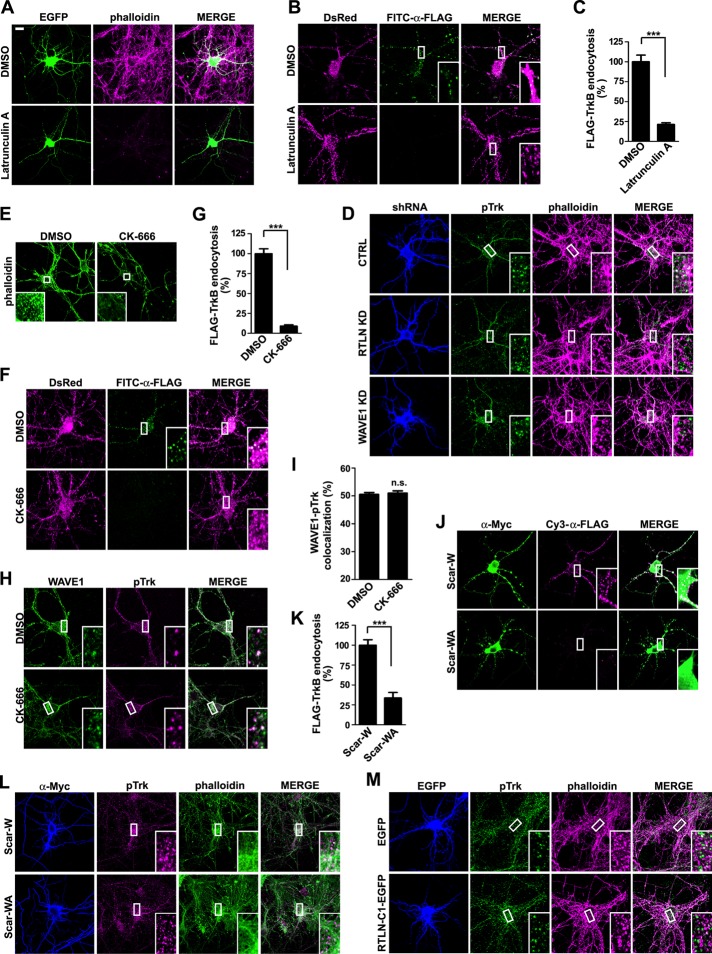
FIGURE 8:
Retrolinkin regulates BDNF-TrkB endocytosis through WAVE1-activated actin polymerization. (A) Hippocampal neurons treated with DMSO (vehicle control) or latrunculin A (1 μM) for 1 h were stained with Alexa Fluor 568–conjugated phalloidin to monitor F-actin content. EGFP serves as volume marker. (B) Hippocampal neurons cotransfected with DsRed and FLAG-TrkB–expressing constructs were treated with DMSO or latrunculin A (1 μM) for 1 h, rinsed with MEM, and stimulated with BDNF (25 ng/ml) for 30 min. Internalized FLAG-TrkB was labeled with FITC–α-FLAG and analyzed by confocal microscopy. (C) Quantification of TrkB endocytosis in neurons in B by measuring mean fluorescence intensity of FLAG-TrkB puncta per cell (35 neurons from two independent experiments). (D) Hippocampal neurons transfected with construct expressing shRNA were starved for 2 h, stimulated with BDNF for 2 min, fixed, and stained with anti-pTrk and Alexa Fluor 568–conjugated phalloidin. Representative confocal images. (E) Hippocampal neurons treated with DMSO or CK-666 (200 μM) for 2 h were stained with Alexa Fluor 488–conjugated phalloidin to monitor F-actin content. (F) Hippocampal neurons cotransfected with DsRed and FLAG-TrkB–expressing constructs were treated with DMSO or CK-666 (200 μM) for 2 h, rinsed with MEM, and stimulated with BDNF (25 ng/ml) for 30 min. Internalized FLAG-TrkB was labeled with FITC–α-FLAG and analyzed by confocal microscopy. (G) Quantification of TrkB endocytosis in neurons in F by measuring mean fluorescence intensity of FLAG-TrkB puncta per cell (42 neurons from three independent experiments). (H) Hippocampal neurons were treated with DMSO or CK-666 (200 μM) for 2 h, rinsed with MEM, and stimulated with BDNF (25 ng/ml) for 2 min and immunostained with antibodies against WAVE1 and pTrk. (I) Quantification of the colocalization between WAVE1 and pTrk in H. Thirty neurons from two independent experiments were analyzed. (J) Hippocampal neurons were cotransfected with expression constructs for FLAG-TrkB and Myc-tagged Scar-W or Scar-WA at DIV4 and treated with BDNF (25 ng/ml) for 30 min at DIV8. Internalized FLAG-TrkB was labeled with Cy3-α-FLAG. Scar-W and Scar-WA were stained with anti-Myc antibody. Scar-W does not bind to Arp2/3 and serves as negative control for Scar-WA. (K) Quantification of Flag-TrkB endocytosis in neurons in J by measuring mean fluorescence intensity of FLAG-TrkB puncta per neuron (25 neurons from two independent experiments). (L) Hippocampal neurons transfected with construct expressing Myc-tagged Scar-W or Scar-WA were starved for 2 h, stimulated with BDNF for 2 min, fixed, and stained with anti-pTrk and Alexa Fluor 568–conjugated phalloidin. Representative confocal images. (M) Hippocampal neurons transfected with construct expressing EGFP or RTLN-C1-EGFP were starved for 2 h, stimulated with BDNF for 2 min, fixed, and stained with anti-pTrk and Alexa Fluor 568–conjugated phalloidin. Representative confocal images. Data represent mean ± SEM. ***p < 0.001, n.s., not significant. Scale bar, 10 μm.

