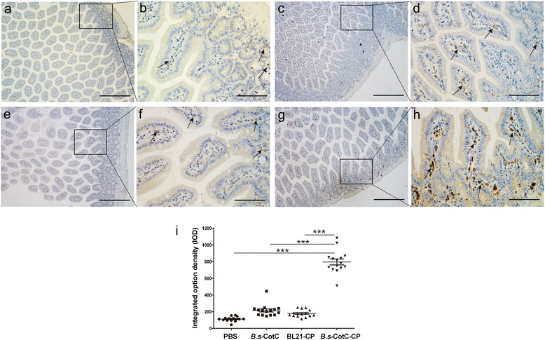
Fig. 6.

Immunohistochemistry analysis of IgA-secreting cells in the intestinal epithelium of orally immunized mice. IgA-secreting cells were stained dark brown. The jejuna (approximately 5–7 mm) of each group were isolated and submitted to immunohistochemical staining at week 4. Panels (a) and (b) represent PBS-treated mice. Panels (c) and (d) represent B.s-CotC orally administered mice. Panels (e) and (f) represent BL21-CsCP gavaged mice. Panels (g) and (h) represent mice orally administered with spores expressing CotC-CsCP. Scale-bars: a, c, e, g, 200 μm; b, d, f, h, 50 μm. The arrows indicate IgA-secreting cells. i Integrated option density (IOD) of IgA-secreting cells. ***P < 0.001
