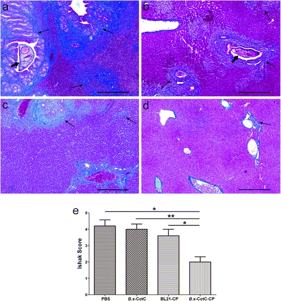
Fig. 9.

Pathological changes of livers from challenge infection mice. Liver sections of each group were stained with Masson’s trichrome. Collagen fibers are shown in blue. Panels a–d show mice orally treated with PBS, B.s-CotC, BL21-CsCP and B.s-CotC-CsCP, respectively. Panel (e) provides the statistic of Ishak scores in liver sections from each mouse. *P < 0.05; **P < 0.01. The thick arrows indicate the adult worms of C. sinensis, and the thin arrows indicate collagen deposition in livers. Scale-bars: 200 μm
