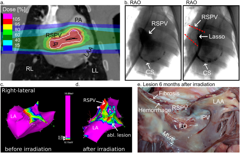
Figure 2. Impact of Carbon Ions on the Right Superior Pulmonary Vein Left Atrial Junction.
(a) Coronal view of a treatment plan for irradiation of the right superior pulmonary vein (RSPV) left atrial (LA) junction. Details as described for Fig. 1a. (b) Right anterior oblique (RAO) view of RSPV venography and RAO of circumferential mapping catheter in the RSPV. Dashed red lines mark projection of RSPV. (c) Right-lateral projection of an endocardial electroanatomical voltage map from the left atrium (LA) and RSPV before irradiation. (d) Right-lateral projection of an electroanatomical voltage map of the LA and RSPV six months after irradiation with 40 Gy carbon ions. Voltage as in legend indicated; voltages >0.7 mV are depicted in magenta and voltage <0.2 mV in red. Other colors mark voltages in-between. (e) Macroscopic lesion outcome at the RSPV-LA junction. The RSPV is opened at its ostium at 12o’clock. Appreciate the macroscopically evident lesion with local hemorrhage and fibrosis. CS = multielectrode catheter in the coronary sinus. Circ. = the circumferential mapping catheter. Turquois dots = double potential. White dots = fragmented signals/area of slow conduction. FO = Transseptal puncture side in the fossa ovalis; LAA = left atrial appendage; MVA = Mitral valve annulus; RSPV = right superior pulmonary vein.

