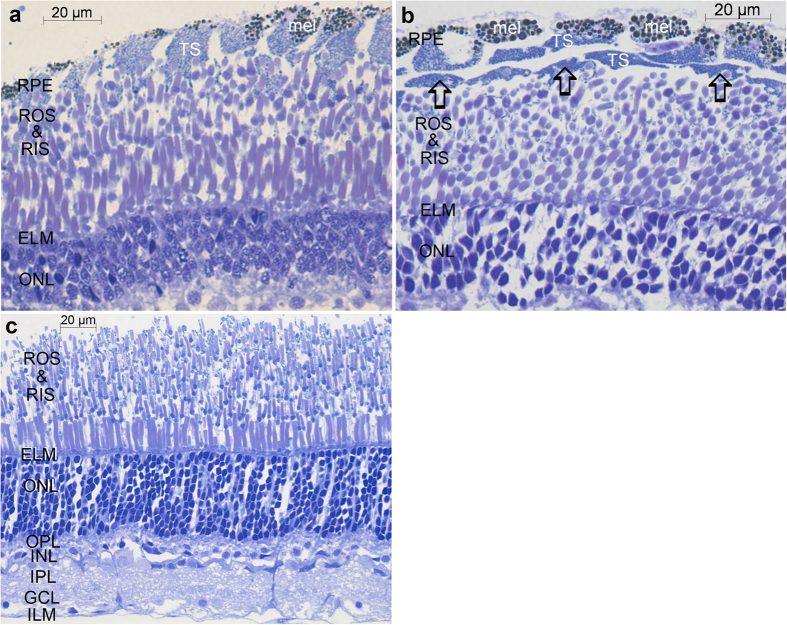
Figure 1. Transverse toluidine blue stained light microscopic sections of the Malacosteus niger retina.
(a,b) show sections of outer retina taken from peripheral regions encompassing the outer nuclear layer (ONL) to the retinal pigment epithelium (RPE). Rod outer segments (ROS) and more darkly stained inner segments (RIS) form several superimposed tiers. Section (a) is cut relatively radially and the RPE cells appear as a single columnar layer with brown melanin pigmentation (mel) sclerally and lighter tapetal spheres (TS) vitreal to the melanin. In (b) the section is cut more obliquely and the RPE thus appears to form 2–3 discrete layers. The arrows indicate oblique sections through the vitreal region of the RPE cells containing the tapetal spheres which, due to the plane of section, appear to be separate from the more scleral part of the RPE cell. ELM–external limiting membrane. (c) is a section of the entire retina taken from more central regions. Here, unlike in the peripheral retina, the photoreceptors nearest the external limiting membrane (ELM) have larger outer segments (ROS) than more scleral rods. There are also more tiers of photoreceptors than peripherally. OPL–outer plexiform layer; INL–inner nuclear layer; IPL–inner plexiform layer; GCL–ganglion cell layer; ILM–internal limiting membrane. In this section the retinal pigment epithelium has become detached.

