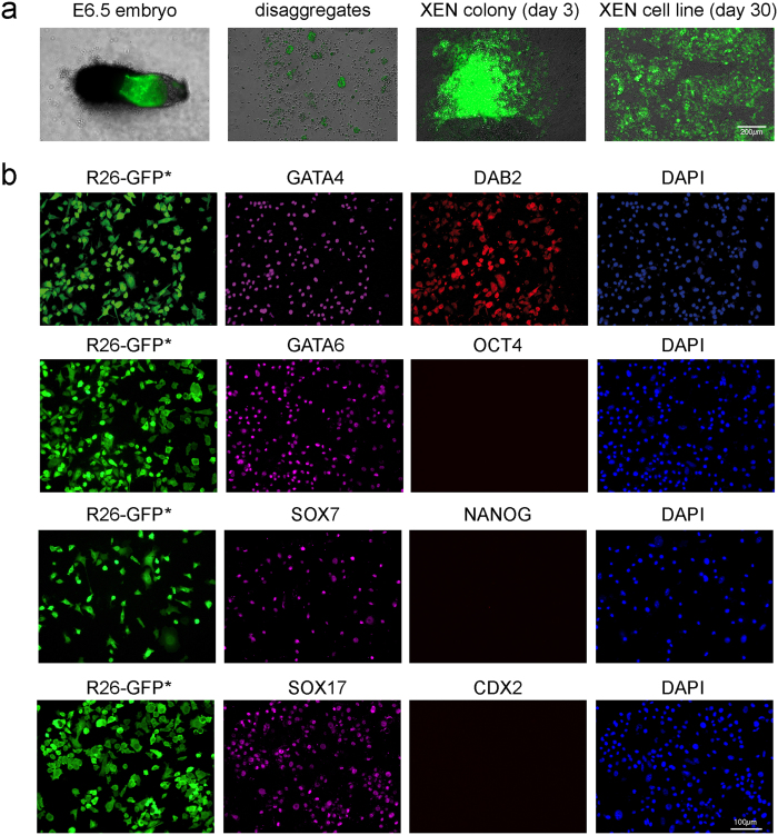
Figure 3. Derivation of post-XEN cell lines from disaggregates of E6.5 embryos.
(a) R26-tauGFP41 × Sox17-Cre E6.5 embryo and post-XEN cell line X-E6.5-Z0617-2. From left to right: whole E6.5 embryo, disaggregated embryo, XEN-like colony expressing GFP on day 3 of culture, and established post-XEN cell line on day 30. Intrinsic green fluorescence of GFP. (b) Fluorescence analysis of post-XEN cell line X-E6.5-Z0617-5. First column: intrinsic (indicated with an asterisk after GFP) green fluorescence of GFP expressed from the ROSA26 locus after activation by Cre recombinase that is expressed from the gene-targeted Sox17 locus. Second column: cells are immunoreactive (magenta) for XEN markers GATA4, GATA6, SOX7, and SOX17. Third column: cells are immunoreactive for XEN marker DAB2, but negative for ES cell markers OCT4 and NANOG, and negative for TS cell marker CDX2. Fourth column: DAPI (blue).

