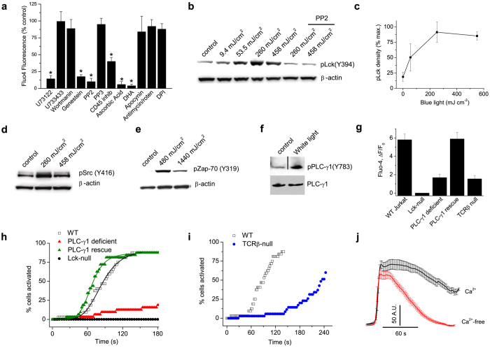
Figure 3. Light activates a Lck-Phospholipase C-γ1 pathway.
(a) Summary of Ca2+ responses evoked by blue light (4.7 mW cm−2, 450 mJ cm−2) in Jurkat cells pretreated (see Methods) with U73122 (500 nM), U733433 (5 μM), wortmanin (1 μM), genestein (100 μM), PP2 (3 μM), PP3 (3 μM), CD45 inhibitor (1 μM), ascorbic acid (10 mM), DHA (docosahexaenoic acid, 5 μM), apocynin (100 μM), antimycin/rotenone/oligomycin (20: 20:5 μM), DPI (diphenyliodonium, 50 μM) (n = 30–40 cells per group in triplicate), *P < 0.001. (b and c) Blue light dose-dependently increases phosphorylation of Lck(Y394) in Jurkat cells detected with a generic pSrc (Y416) antibody (see Supplementary Fig. 3), data are mean of 3 experiments. The Src inhibitor, PP2, blocks the increase in pY394 demonstrating that light triggers trans autophosphorylation of Lck. (d) Blue light increases pSrc in murine CD3+ T cells. (e) Blue light triggers phosphorylation of ZAP-70 (Y319) in Jurkat cells. (f) White light (13.7 J cm−2) stimulates tyrosine phosphorylation (Y783) of PLC-γ1. Note that the immunoblots are cropped and the full blots are shown in Supplementary Fig. 9. (g–i) Mean changes in Fluo4 fluorescence and cumulative activation plots in response to blue light for wild-type Jurkat cells, PLC-γ1-deficient Jurkat T cells (Jgamma1), Jgamma1 cells stably expressing PLC-γ1 (JgammaWT), Lck-deficient Jurkat cells (JCam1.6) and cells lacking TCRβ expression (n = 40–120 for each group). (j) Light-evoked Ca2+ responses in control and Ca2+-free media; n = 40–50. The rising phase of responses is aligned to show the time course of decay.

