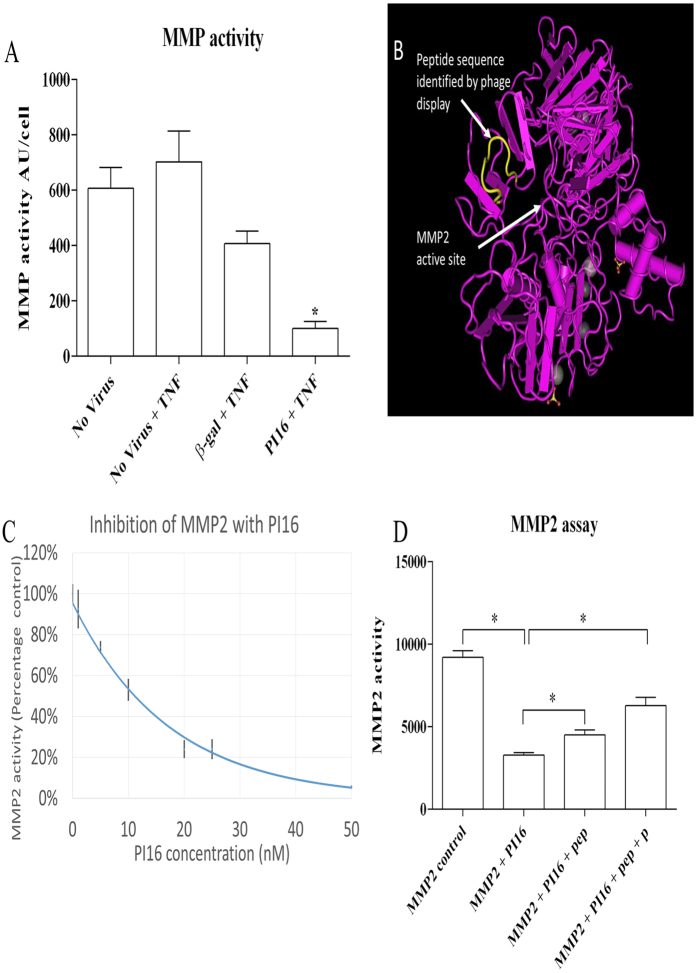
Figure 4. PI16 reduces MMP activity.
(A) Concentrated conditioned media from control or PI16 over-expressing HCEACs was assessed for MMP activity using a quenched-fluorogenic substrate OmniMMP. In comparison to all controls, PI16 conditioned media showed the lowest MMP activity (*p < 0.05, n = 5). (B) Phage display identified a peptide containing the sequence TGPRSDGF, with high homology to amino acids 250–256 of MMP2 TG-RSDGF, corresponding to a peptide loop situated above the active site of MMP2. (C) Recombinant MMP2 (5.5 nM) was incubated with increasing concentrations of PI16 for 30 minutes before the addition of OmniMMP. Relative fluorescence 30 minutes after addition of OmniMMP is presented. Graph combines data from 3 separate preparations of recombinant PI16. Concentrations above 10 nM resulted in a significant reduction of MMP2 activity (P < 0.05, n = 3–23). (D) Inclusion of peptides TGRSDGF (pep) or TGPRSDGF (pep + P) (10 μM), with 10 nM PI16 reduced the inhibition of MMP2 (*p < 0.05 compared to all other conditions, n = 3).

