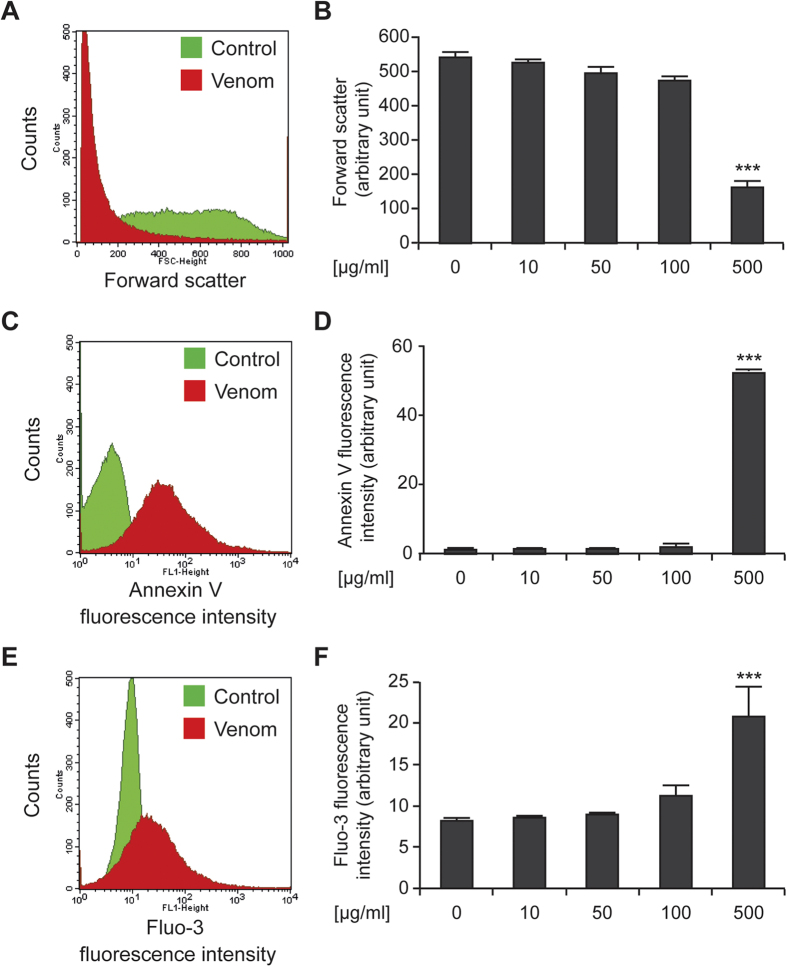
Figure 1. Trachinus vipera venom induces eryptosis.
(A,B) Effect of venom on the erythrocytes size. Erythrocytes were maintained in Ringer solution followed by treatment or not for 48 h with 10 to 500 μg/ml of venom. The forward scatter of erythrocytes was estimated by flow cytometry. (A) Illustrates representative dot plots (control was labeled in green and 500 μg/ml venom in red), while (B) report quantitative data. Data are reported as means ± SEM (n = 9). (C,D) Effect on phosphatidylserine exposure. Erythrocytes (control and treated ones) was labeled with annexin-V for the assessment of apoptosis-associated parameters (phosphatidylserine exposure). (B) Illustrates representative dot plots (control was labeled in green and 500 μg/ml venom in red), while (C) report quantitative data. Data are reported as means ± SEM (n = 9). (E,F) Effect of venom on erythrocyte Ca2+ activity. Erythrocytes (control and treated ones) was labeled with Fluo3-AM for the assessment of erythrocyte cytosolic Ca2+ concentration. (E) Illustrates representative dot plots (control was labeled in green and 500 μg/ml venom in red), while (F) report quantitative data. Data are reported as means ± SEM (n = 9). ***(p < 0.001) indicate significant difference as compared with non-treated erythrocyte (ANOVA).

