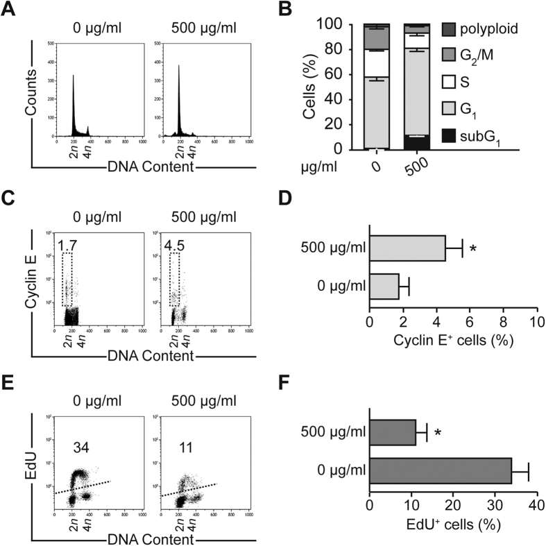
Figure 4. Trachinus vipera venom induces accumulation in G1 phase of cell cycle and stops DNA synthesis.
(A,B) Cell cycle analysis. Human colon carcinoma HCT116 cells were exposed to 500 μg/ml Trachinus vipera venom for 72 h followed by the cytofluorometric assessment of cell cycle distribution. (A,B) report representative cell cycle distributions and quantitative data (mean ± SEM; n = 3). (C,D) G1 phase staining. HCT116 cells were cultured in the presence of venom for 72 h, fixed and then labeled with an antibody recognizing cyclin E, which accumulates essentially in the G1 phase. Cells were acquired using cytofluorometry. Representative scatter plots (C) and quantitative results (D) (mean ± SEM; n = 3) are shown. (E,F) DNA synthesis evaluation. HCT116 cells were cultured in the presence of 500 μg/ml Trachinus vipera venom and after 72 h labeled with the thymidine analog EdU, which selectively stains cells in the S phase of the cell cycle. Cell were fixed and acquired by cytofluorometry to evaluate the level of EdU incorporation. Representative scatter plots (E) and quantitative results (F) (mean ± SEM; n = 3) are shown. Numbers indicate the percentage of cells found in each quadrant. *p < 0.05 (Student’s t-test), as compared with non-treated cells.

