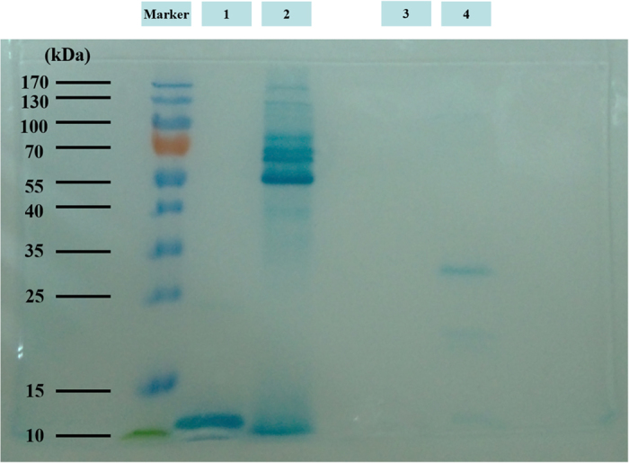
Figure 4. LDS-PAGE of EPS of the three bacterial strains and bovine heart cytochrome c with heme staining.

1: bovine heart cytochrome c, 2: EPS of S. oneidensis, 3: EPS of E. coli, and 4: EPS of P. putida.

1: bovine heart cytochrome c, 2: EPS of S. oneidensis, 3: EPS of E. coli, and 4: EPS of P. putida.