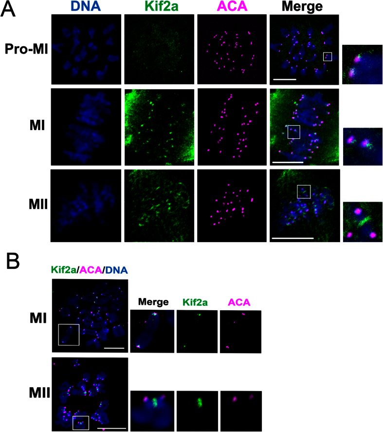
Figure 2. Localization of Kif2a on chromosomes.
(A) Co-localization of Kif2a with ACA at Pro-M I,M I, and M II stages. Oocytes cultured to 4 h (Pro-M I), 8 h (M I), and 12 h (M II) were stained for Kif2a (green), ACA (pink), and DNA (blue). Bar = 10 μm. (B) Colocalization of Kif2a and ACA at M I and M II stages. Oocytes were cultured to M I and M II stages, then chromosomes were spread and stained with Kif2a (green), ACA (pink), and DNA (blue). Magnifications of the boxed regions were displayed on the right of the main panel. Bar = 10 μm.

