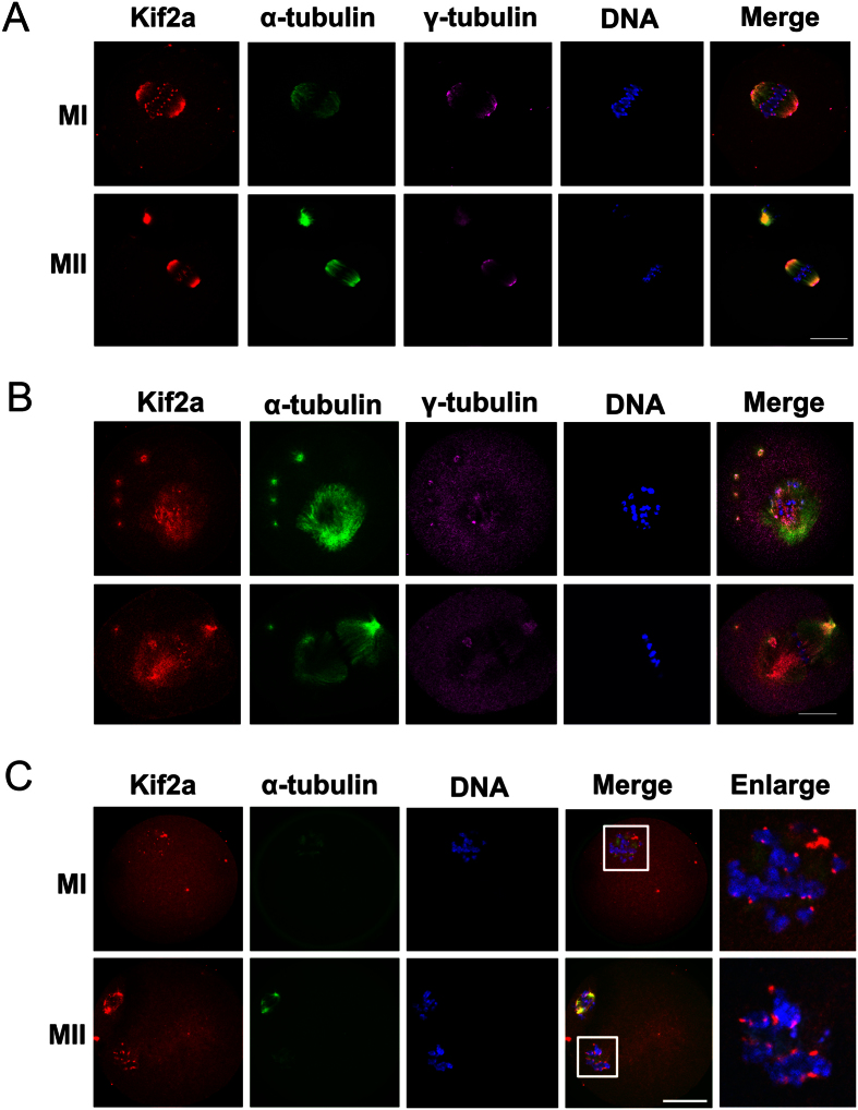
Figure 3. Localization of Kif2a in mouse oocytes treated with spindle perturbing agents, taxol or nocodazole.
(A) Oocytes were collected at 8 h or 12 h of culture in M2 medium containing DMSO stock. And then triple stained with antibodies against Kif2a, γ-tubulin, and anti-α-tubulin-FITC. (B) Oocytes at the M I stage were incubated in M2 medium containing 10 μm taxol for 45 min and then triple stained with antibodies against Kif2a, γ-tubulin, and anti-α-tubulin-FITC. Kif2a was localized at the enlarged spindle, congressed spindle poles, centromeres, and cytoplasmic asters. (C) Oocytes at M I and M II stages were incubated with 20 μg/ml nocodazole in M2 medium for 15 min and then double stained for Kif2a and α-tubulin. Red, Kif2a; green, α-tubulin; pink, γ-tubulin; blue, chromatin; Magnifications of the boxed regions were displayed on the right of the main panel. Bar = 20 μm.

