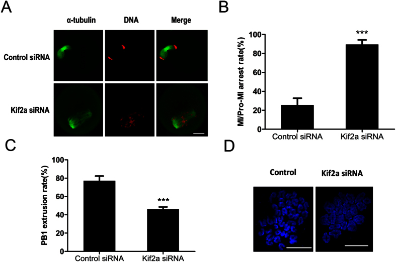
Figure 6. Depletion of Kif2a arrests oocytes at the Pro-MI/MI stage and decreases PB1 extrusion.
Oocytes injected with Kif2a siRNA or control siRNA were cultured in fresh M2 medium for 10 h or 12 h, after being incubated in M2 medium containing 100 μm IBMX for 24 h. (A) Oocytes cultured for 10 h after IBMX washout in the Kif2a-depletion group were arrested at the Pro-MI/MI stage, whereas oocytes in the control group reached the AI stage. Injected oocytes were stained with α-tubulin (green); DNA (red). Bar = 20 μm. (B) The rate of Pro-MI/MI cultured oocytes was recorded in the Kif2a knockdown group and the control group at 10 h of culture. Data are presented as Mean ± SEM of 3 independent experiments (***P < 0.001). (C) Percentages of PB1 extrusion of oocytes cultured for 12 h in the Kif2a knockdown group and control group. Data are presented as Mean ± SEM of 3 independent experiments (***P < 0.001). (D) Chromosome spreading showed failure of homologous chromosome segregation in the Kif2a knockdown group after culture for 12 h. The percentage for bivalents was 35/46 in the knockdown group and 16/56 in the control group. Bar = 20 μm.

