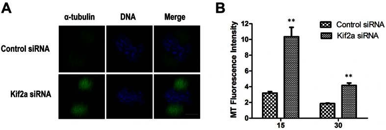
Figure 8. Kif2a controls spindle dynamics in mouse oocytes.
Oocytes in MI stages were incubated at 4 °C for 15 min or 30 min to selectively depolymerize non-kinetochore microtubules and fixed, then stained for α-tubulin and DNA. (A) Single projection of z sections spanning the entire spindle width of the representative control group or Kif2a knockdown group are shown. Images for α-tubulin were obtained under a constant exposure parameter; green, α-tubulin; blue, DNA. Bar = 20 μm. (B) The fluorescence intensity of α-tubulin on spindles of the Kif2a knockdown group at the 15 min and 30 min time points were quantified and normalized to their respective control groups. (n = 10 cells for each quantification). (**P < 0.01).

