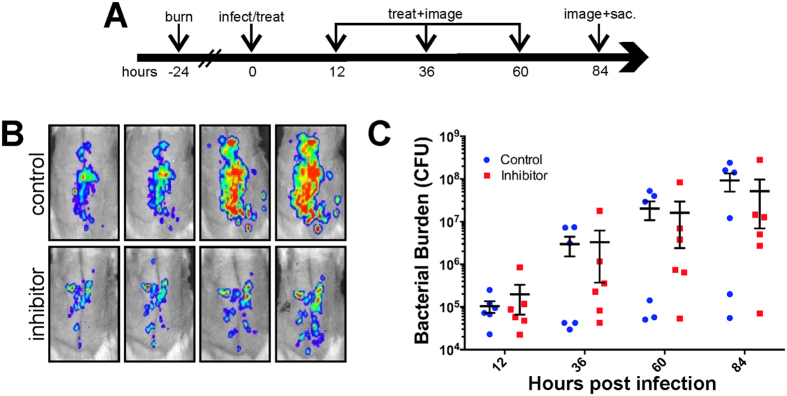
Figure 3. Inhibition of P. aeruginosa in unexcised burns.
Schematic of infection and dosage regimen (A). Representative bioluminescence images of MAM7 inhibitor treated (bottom row) and control bead treated (upper row) rats infected with P. aeruginosa (B). Quantification of bacterial burden by IVIS photon flux analysis (C).

