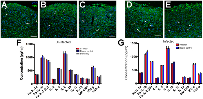
Figure 6. Neutrophil infiltration and vascularization of burned skin is unimpaired by GST-MAM7 beads.
Confocal micrographs of burned skin (as in boxed area depicted in Fig. 1A) were used to analyze neutrophil infiltration (bright green fluorescent clumps) and vascularization (observed through green channel background) for the following samples: (A) burned skin only, (B) burned skin with GST-beads, (C) burned skin with GST-MAM7-beads, (D) burned skin with GST-beads + P. aeruginosa, and (E) burned skin with GST-MAM7-beads + P. aeruginosa. Arrows point to blood vessels. Scale bar equals to 150 μm. (F) Serum cytokine levels of burned only (grey), burned inhibitor (red) or burned control (blue) treated uninfected animals. (G) Serum cytokine levels of inhibitor (red) or control (blue) treated animals with P. aeruginosa infected burns. Data presented are means ± stdev (n = 6 for inhibitor and bead treated groups, n = 5 for burn only group, each sample measured in triplicate against cytokine standards). All samples were taken post-mortem (6 d.p.i.).

