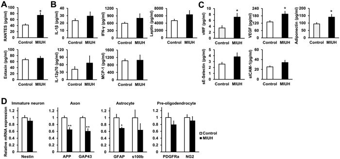
Figure 6. Levels of inflammatory cytokines and chemokines and ischemia-related proteins in the placenta on E20.
The levels of several inflammatory and ischemia-related factors were measured via a multiplex assay. (A) Inflammatory chemokines. RANTES levels were significantly increased, whereas eotaxin levels did not change in the placenta 3 days after coil stenosis. (B) Inflammatory cytokines. Cytokine levels did not significantly change in the placenta following mild intrauterine hypoperfusion (MIUH) on E17, although all inflammatory cytokines showed increasing trends. n = 11 in the control group and n = 12 in the MIUH group. (C) Ischemia-related proteins. The levels of vWF, VEGF, and adiponectin were significantly increased in the placenta following MIUH. n = 13 in the control group and n = 14 in the MIUH group. *P < 0.05, vs. the control group. (D) mRNA expression levels of an immature neuron maker (nestin), axonal makers (APP and GAP43), astrocyte makers (GFAP and S100β), and oligodendrocyte makers (PDGFRα and NG2) in the whole brain (n = 5 in each group). β-actin was used as an internal control for gene expression. The mRNA expression levels of APP, GAP-43 and GFAP in brains on E20 were significantly decreased following MIUH. *P < 0.05, **P < 0.01 vs. the control group. E, Embryonic day.

