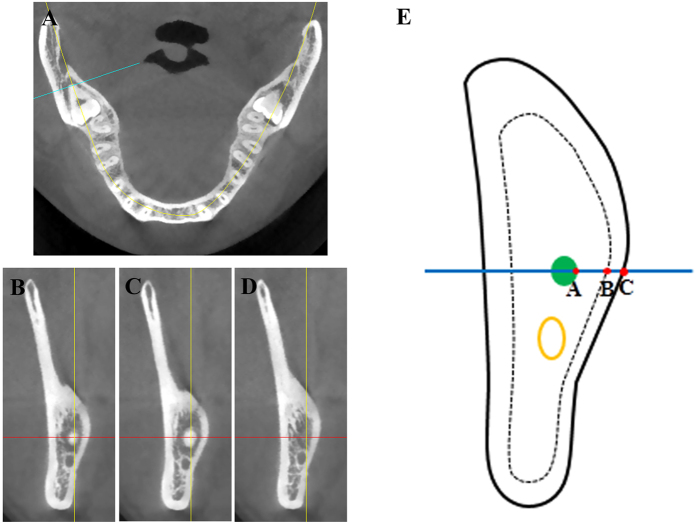
Figure 2. Selection of cross-sectional CBCT images when root apex of impacted mandibular third molar was located at its mostly distal slice and subsequent relevant measurements.
(A) The root apex of impacted mandibular third molar which was localized most distally was initially identified on the axial CBCT image. (B–D) Starting from the (A) image, the CBCT slice was scrolled distally or mesially. When the root tip was enlarged (C) or disappeared (D) on the next slices after the image was adjusted mesially or distally (0.1 mm). the present image (B) on the cross-sectional plane was further selected for various measurements and spatial analyses. (E) Schematic illustration of the measurements. Point A was identified as the most lingual site of root apex. A horizontal line through point A was automatically generated which was intersected with the inner (Point B) and outer surface (Point C) of lingual plate, respectively. The measurements such as thickness of lingual plate (B–C) and distance between root apex and the outer surface of lingual plate (A–C) were performed using the digital ruler provided by the software Simplant.

