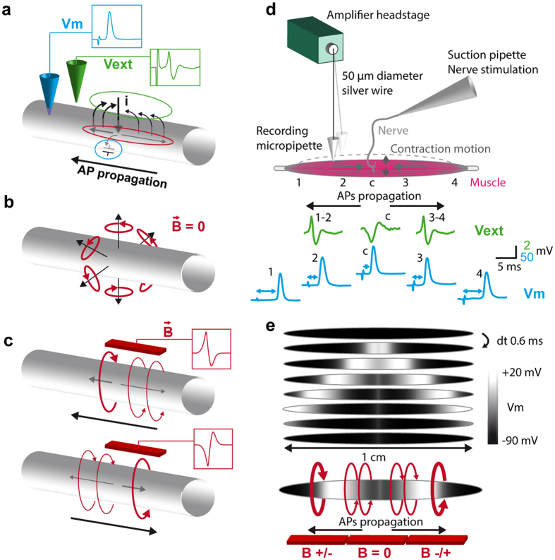
Figure 1. Electrophysiology.
(a) Transmembrane and axial currents in an active zone of an excitable cable. Charge of the membrane capacitor is the quantity measured with an intracellular pipette (blue circle). An extracellular electrode measures the effect of the transmembrane currents on the external potential (green circle). Axial currents (red circle) are not directly measured with classical electrophysiology. (b) Theoretical magnetic fields associated to transmembrane currents regularly distributed along the circumference of a cable. Seen from a certain distance, contributions of the transmembrane currents to the net magnetic field cancel each other. (c) Theoretical magnetic field associated to axial currents flowing in front and back of an action-potential in an excitable cable. Sign of the magnetic field depends on the current direction. (d) Scheme depicts the floating electrode technique used for intra and extracellular recordings in the contracting muscle. The extreme tip of a glass micropipette is used as electrode, and hangs at the extremity of a free moving silver wire connected by its opposite end to the amplifier headstage. The absence of pipette holder allows the free moving of the system and the intracellular recordings during contraction. Green traces represent the extracellular potential measured in the synaptic region, and on both sides of the muscle, when the nerve is electrically stimulated. Blue traces represent the intracellular recording of the muscle AP, in the synaptic region and in different positions on both sides of the central region. (e) Gray scale representation of the simulated membrane potential at different times showing the internal gradients at the front and the back of the two active zones. Bottom scheme represents the expected magnetic fields associated to the muscle AP.

