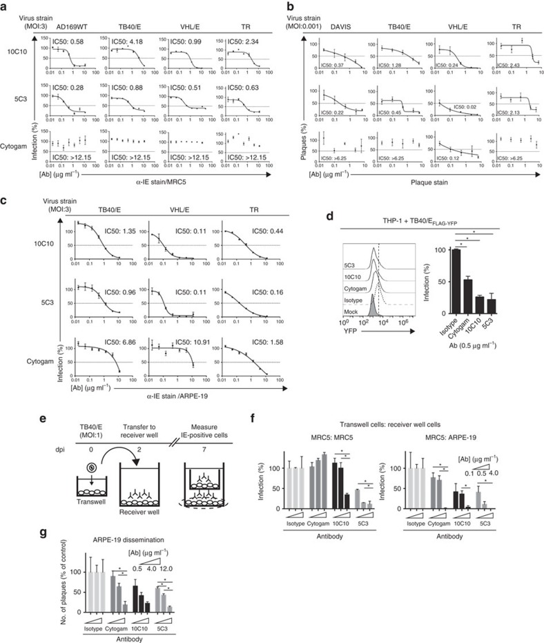
Figure 4. Examination of the potency and versatility of anti-gH mAbs in epithelial cells.
(a) Cytogam and mAbs 10C10 and 5C3 were pre-incubated with CMV strains AD169, TB40/E, VHL/E and TR (0.01–12 μg ml−1), and subsequent infection levels of MRC5 cells was analyzed by immunostaining with anti-Immediate Early (IE) gene product (α-IEAlexa488) antibody. Experiments were performed in technical triplicate. (b) Cytogam and mAbs 10C10 and 5C3 were pre-incubated with CMV strains DAVIS, TB40/E, VHL/E and TR (0.025–6.25 μg ml−1), and subsequent infection levels of MRC5 cells was analyzed by plaque assay. Data represents averages from three experiments performed in duplicate. (c) Cytogam and mAbs 10C10 and 5C3 were pre-incubated with CMV strains TB40/E, VHL/E and TR (0.01–12 μg ml−1), and subsequent infection levels of human retinal pigment epithelial cells (ARPE-19) was measured by anti-IE staining. Experiments were performed in technical triplicate. Non-linear regression analysis was performed and the half maximal inhibitory concentration (IC50) was calculated for all antibodies under all conditions. (d) Cytogam and mAbs 10C10 and 5C3 were pre-incubated with the fluorescent CMV strain TB40/EFLAG-YFP (0.5 μg ml−1) before infection of THP-1 cells (MOI: 3) and fluorescence levels were analyzed by flow cytometry at 4 dpi. Experiments were performed in technical triplicate. (e) MRC5 cells were seeded on a transwell insert with 3 μm pore and infected (MOI:1) with TB40/E. At 2 dpi the transwell insert was transferred to a receiver well containing an isotype control, Cytogam or mAbs 10C10 and 5C3 (0.1–4 μg ml−1) and at 7 dpi the cells from the receiver layer were analyzed by α-IEAlexa488 immunostain. (f) MRC5 (left panel) and ARPE-19 (right panel) cells from the transwell infection experiment were analyzed for infection levels by α-IEAlexa488 immunostain. Experiments were performed in technical triplicate. (g) Infection levels of ARPE-19 cells infected with TB40/E (MOI:0.1) and exposed to an isotype control, Cytogam or mAbs 10C10 and 5C3 (0.5–12 μg ml−1) over a 10 day period were analyzed by α-IEAlexa488 immunostain. Experiments were performed in technical triplicate. S.d. is depicted for all experiments. *P<0.05 (Student's two-tailed t test).

