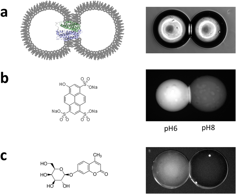
Figure 1. Assembling a Droplet Interface Bilayer system for the transport of small sugars through LacY.
(a) Schematic of a DIB with LacY incorporated (left) with a brightfield image of two droplets forming the bilayer at the point of contact (right panel). (b) The pH sensitive dye pyranine (left) was loaded in two droplets at a concentration of 50 μM dye in 50 mM sodium phosphate buffer of two different pH. The image shown on the right was taken after 1 hour using a fluorescent microscope. (c) The fluorescent sugar MUG (left) is a substrate of LacY. A DIB was formed from DOPC lipids (right panel). The left hand side droplet was loaded with 50 μM MUG while the right hand side droplet contained only buffer. The image shown was taken after 1 hour.

