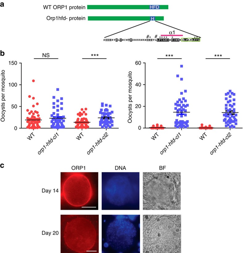
Figure 5. The HFD is necessary for ORP1 function.
(a) Schematic depiction of the construct encoding ORP1 in which the α2 to αC helices (aa 797–860) of the HFD were deleted leaving the epitope for the ORP1 antibody intact (green box). (b) Oocyst load in mosquitoes infected with orp1-hfd−compared with WT at day 11 (left) and day 21 p.b.f. (right). Pooled data from two independent experiments of each clone. NS, nonsignificant, ***P<0.0001, Mann–Whitney test. The complete data set is presented in Supplementary Fig. 9 and Supplementary Table 1. Error bars denote s.e.m. (c) ORP1 lacking the HFD is localized at the oocyst capsule at both day 14 and 20 p.b.f. Sporozoites are visible inside the oocysts. Nuclei stained with 4,6-diamidino-2-phenylindole (DAPI; blue) and the bright field (BF) image. Scale bars, 20 μm.

