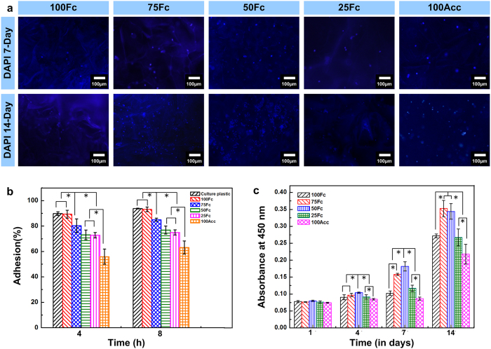
Figure 7. Adhesion, morphology and proliferation of L929 cells on the scaffolds.
(a) The cells growing on the scaffolds after 7 and 14-day culture were stained with DAPI for nuclei (blue). The fluorescence of DAPI was more intense than the background fluorescence from scaffolds, thus providing the sufficient contrast for imaging. Scale bars, 100 μm. (b) The adhesion of L929 cells on the surface of the culture dishes and the scaffolds (n = 3 per group). (c) The cell viability on the scaffolds after 7 and 14-day culture (n = 3 per group). * denotes statistically significant differences. (n = 6 per group, p < 0.05).

